Thje structure of part of a DNA.(double helix)
Deoxyribonucleic acid or DNA, is a ncleic acid that contains the genetic instructions used in the development and functioning of all known living organisms . The main role of DNA molecules is the long-term storage of information. DNA is often compared to a set of blueprints, like a recipe or a code, since it contains the instructions needed to construct other components of cells, such as proteins and RNA molecules. The DNA segments that carry this genetic information are called genes, but other DNA sequences have structural purposes, or are involved in regulating the use of the genetic information.
DNA consists of two long polymers of simple units called nucleotides, with backbones made of sugars and phosphate groups joined by ester bonds. These two strands run in opposite directions to each other and are therefore anti-parallel. Attached to each sugar is one of four types of molecules called bases. It is the sequence of these four bases along the backbone that encodes information. This information is read using the genetic code, which specifies the sequence of the amino acids within proteins. The code is read by copying stretches of DNA into the related nucleic acid RNA, in a process called transcription.
Within cells, DNA is organized into long structures called chromosomes. These chromosomes are duplicated before cells divide, in a process called DNA replication. Eukaryotic organisms (animals, plants, fungi, and protists) store most of their DNA inside the cell nucleus and some of their DNA IN organelles, such as mitochondria or chloroplasts. In contrast, prokaryotes (bacteria and archaea) store their DNA only in the cytoplasm. Within the chromosomes, chromatin proteins such as histones compact and organize DNA. These compact structures guide the interactins between DNA and other proteins, helping control which parts of the DNA are transcribed.
DNA SPECIAL
Wednesday, 23 February 2011
Sunday, 20 February 2011
DNA Structure
DNA Structure
Biology the DNA is a beautiful things to learn about.
A very good tables to show DNA
Same Living things the world.
Legend:
Illustration of the double helical structure of the DNA molecule.
The structure of DNA is illustrated by a right handed double helix, with about 10 nucleotide pairs per helical turn. Each spiral strand, composed of a sugar phosphate backbone and attached bases, is connected to a complementary strand by hydrogen bonding (non- covalent) between paired bases, adenine (A) with thymine (T) and guanine (G) with cytosine (C).
Adenine and thymine are connected by two hydrogen bonds (non-covalent) while guanine and cytosine are connected by three.
This structure was first described by James Watson and Francis Crick in 1953.
Saturday, 19 February 2011
DNA Structure
Introduction to DNA Structure
A Molecular Graphics companion to and Introductory Course in Biology or Biochemistry.
Contents
Components of DNA
DNA is a polymer. The monomer units of DNA are nucleotides, and the polymer is known as a "polynucleotide." Each nucleotide consists of a 5-carbon sugar (deoxyribose), a nitrogen containing base attached to the sugar, and a phosphate group. There are four different types of nucleotides found in DNA, differing only in the nitrogenous base. The four nucleotides are given one letter abbreviations as shorthand for the four bases.
Adenine and guanine are purines. Purines are the larger of the two types of bases found in DNA. Structures are shown below.
Structure of A and G
The 9 atoms that make up the fused rings (5 carbon, 4 nitrogen) are numbered 1-9. All ring atoms lie in the same plane.
Pyrimidine Bases
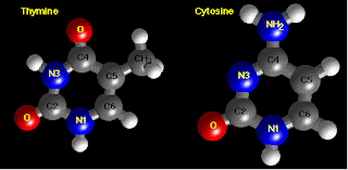
Cytosine and thymine are pyrimidines. The 6 stoms (4 carbon, 2 nitrogen) are numbered 1-6. Like purines, all pyrimidine ring atoms lie in the same plane.
Structure of C and T
Deoxyribose Sugar
The deoxyribose sugar of the DNA backbone has 5 carbons and 3 oxygens. The carbon atoms are numbered 1', 2', 3', 4', and 5', to distinguish from the numbering of the atoms of the purine and pyrmidine rings. The hydroxyl groups on the 5'- and 3'- carbons link to the phosphate groups to from the DNA backbone. Deoxyribose lacks an hydroxyl group at the 2'-position when compared to ribose, the sugar component of RNA.
Structure of deoxyribose
Nucleosides
A nucleoside is one of the four DNA bases covalently attached to the C1' position of a sugar. The sugar in deoxynucleosides is 2'-deoxyribose. The Sugar in ribonucleosides is ribose. Nucleosides differ from nucleotides in that they lack phosphate groups. The four different nucleosides of DNA are deoxyadenosin (dA),
Structure of dA
In dA and dG, there is an "N-glycoside" bond between the sugar C1' and N9 of the purine
Nucleotides
A nucleotide is a nucleoside with one or more phosphate groups covalently attached to the 3' - and/ 5'-hydroxyl group(s).
DNA Backbone
The DNA backbone is a polymer with an alternating sugar-phosphate sequence. The deoxyribose sugars are joined at both the 3' -hydroxyl and 5' -hydroxyl groups to phosphate groups in ester links, also known as "phosphdiester" bonds.
Example of DNA Backbone: 5'-d(CGAAT):
Features of the 5'-d(CGAAT) structure:
DNA is a normally double stranded macromolecule. Two polynucleotide chains, held together by weak thermodynamic forces, from a DNA molecule.
Structure of DNA Double Helix
Features of the DNA Double Helix
Within the DNA double helix, A forms 2 hydrogen bonds with T on the opposite strands, and G forms 3 hyrdorgen bonds with C on the opposite strand.
Example of dA-dT base pair as found within DNA double helix
Example of dG-dC base pair as found within DNA double helix
The helix axis is most apparent from a view directly down the axis. The sugar-phosphate backbone is on the outside of the helix where the polar phosphate groups (red and yellow atoms) can interact with the polar environment. The nitrogen (blue atoms) containing bases are inside, stacking perpendicular to the helix axis.
View down the helix axis
A Molecular Graphics companion to and Introductory Course in Biology or Biochemistry.
Contents
- Components of DNA
- Purine Bases
- Pyrimidine Bases
- Deoxyribose Sugar
- Nucleosides
- Nucleotides
- Base Pairs
- DNA Backbone
- DNA Double Helix
- DNA Helix Axis
Components of DNA
DNA is a polymer. The monomer units of DNA are nucleotides, and the polymer is known as a "polynucleotide." Each nucleotide consists of a 5-carbon sugar (deoxyribose), a nitrogen containing base attached to the sugar, and a phosphate group. There are four different types of nucleotides found in DNA, differing only in the nitrogenous base. The four nucleotides are given one letter abbreviations as shorthand for the four bases.
- A is for adenine
- G is for guanine
- C is for cytosine
- T is for thymine
Adenine and guanine are purines. Purines are the larger of the two types of bases found in DNA. Structures are shown below.
Structure of A and G
The 9 atoms that make up the fused rings (5 carbon, 4 nitrogen) are numbered 1-9. All ring atoms lie in the same plane.
Pyrimidine Bases
Cytosine and thymine are pyrimidines. The 6 stoms (4 carbon, 2 nitrogen) are numbered 1-6. Like purines, all pyrimidine ring atoms lie in the same plane.
Structure of C and T
Deoxyribose Sugar
The deoxyribose sugar of the DNA backbone has 5 carbons and 3 oxygens. The carbon atoms are numbered 1', 2', 3', 4', and 5', to distinguish from the numbering of the atoms of the purine and pyrmidine rings. The hydroxyl groups on the 5'- and 3'- carbons link to the phosphate groups to from the DNA backbone. Deoxyribose lacks an hydroxyl group at the 2'-position when compared to ribose, the sugar component of RNA.
Structure of deoxyribose
Nucleosides
A nucleoside is one of the four DNA bases covalently attached to the C1' position of a sugar. The sugar in deoxynucleosides is 2'-deoxyribose. The Sugar in ribonucleosides is ribose. Nucleosides differ from nucleotides in that they lack phosphate groups. The four different nucleosides of DNA are deoxyadenosin (dA),
Structure of dA
In dA and dG, there is an "N-glycoside" bond between the sugar C1' and N9 of the purine
Nucleotides
A nucleotide is a nucleoside with one or more phosphate groups covalently attached to the 3' - and/ 5'-hydroxyl group(s).
DNA Backbone
The DNA backbone is a polymer with an alternating sugar-phosphate sequence. The deoxyribose sugars are joined at both the 3' -hydroxyl and 5' -hydroxyl groups to phosphate groups in ester links, also known as "phosphdiester" bonds.
Example of DNA Backbone: 5'-d(CGAAT):
Features of the 5'-d(CGAAT) structure:
- Alternating backbone of deoxyribose and phosphodiester groups
- Chain has a direction (known as polarity), 5'- to 3'- from top to bottom
- Oxygens (red atoms) of phosphates are polar and negatively charged
- A, G, C and T bases can extend away from chain, and stack atop each other
- Bases are hydrophobic
DNA is a normally double stranded macromolecule. Two polynucleotide chains, held together by weak thermodynamic forces, from a DNA molecule.
Structure of DNA Double Helix
Features of the DNA Double Helix
- Two DNA strands form a helical spiral, winding around a helix axis in a right-handed spiral
- The two polynucleotide chains run in opposite directions
- The sugar-phosphate backbones of the two DNA strands wind around the helix axis like the railing of a sprial staircase
- The bases of the individual nucleotides are on the inside of the helix, stacked on top of each other like the steps of a spiral staircase.
Within the DNA double helix, A forms 2 hydrogen bonds with T on the opposite strands, and G forms 3 hyrdorgen bonds with C on the opposite strand.
Example of dA-dT base pair as found within DNA double helix
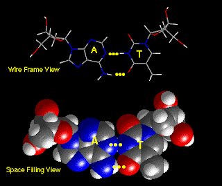
Example of dG-dC base pair as found within DNA double helix
- dA-dT and dG-dC base pairs are the same length, and occupy the same space within the DNA double helix. Therefore the DNA molecule has a uniform diameter.
- dA-dT and dG-dC base pairs can occur in any order within DNA molecules
The helix axis is most apparent from a view directly down the axis. The sugar-phosphate backbone is on the outside of the helix where the polar phosphate groups (red and yellow atoms) can interact with the polar environment. The nitrogen (blue atoms) containing bases are inside, stacking perpendicular to the helix axis.
View down the helix axis
DNA
DNA rarest (deoxyribonucleric acid)
DNA mode identifies - First European Farmers
HIV STRUCTURE CD4+ white blood cell, and the spot on its surface and the spiky blue objects inthe foreground represnt HIV particles.
Human Immunodeficiency Virus- Structure
The proteins gp 120 & gp41 togetber make up the spikes that project from HIV particles while p 17 forms the matrix & p24 forms the core
Assembly, Buddidng & maturation. This electron microscope photo shows newly formed HIV particle budding from a human cell.
DNA, or deoxyriboknucleic acid, is the hereditry material in humans and almost all other organism. Nearly every cell in a person's body has the same DNA. Most DNA is located in the cell nucleus it is called mitochondrial DNA OR mtDNA.
Subscribe to:
Posts (Atom)